Discovering and Treating Vascular Birthmarks
“Take a look at this,” my son’s pediatrician said, as he motioned to two nurses standing outside of the Winnie-the-Pooh themed exam room.
Roughly one week earlier, I had discovered a small, flat, bright red line on the corner of my son’s lower left lip. He was a voracious nurser, so I believed that this line was some sort of nursing blister. I thought it would go away within a few days. But almost a week later, my husband and I noticed no change, so I immediately made an appointment with our pediatrician.
The nurses came into the exam room on this unseasonably cool spring morning and the pediatrician put his gloved finger on my baby’s lip. The three of them looked intently at the red line, as my stomach continued to turn in violent knots. My precious four-week-old lay peacefully on the table, alternately staring with his huge brown eyes at me, the doctor and the Winnie-the-Pooh and Piglet wallpaper, blissfully oblivious to not only what was happening around him, but to his mother’s increasing fears.
“What is it?” I asked, struggling to get the words out.
“It’s a hemangioma – a vascular birthmark,” our pediatrician replied.
This diagnosis initiated a flood of research in the days, weeks and months that lay ahead of our family. But with the help of the Internet and organizations devoted to providing families like ours with the latest vascular birthmark information and treatment resources, the knowledge we gained alleviated our fears.
Vascular Birthmark Statistics
According to the American Academy of Dermatology, 10 percent of the approximate 4 million children born in the U.S. each year will arrive with a vascular birthmark. These birthmarks consist of blood vessels bunched together within the skin. They can appear flat or raised, and be either red or blue in color. There are several different kinds of vascular birthmarks, but the most common are port-wine stains and hemangiomas.
Port-Wine Stains
Port-wine stains are classified as vascular malformations. The Nemours Foundation, based in Wilmington, Del., reports that they occur in about three of every 1,000 births and are diagnosed equally among males and females. Port-wine stains develop when an area of skin doesn’t receive an adequate supply of nerve fibers to assist in keeping the blood vessels contracted. When there is a lack of nerve fibers, capillaries (small blood vessels) continue to expand, allowing a large amount of blood to flow into the blood vessels, which causes a “stain” to appear under the skin. Port-wine stains commonly appear on the face, but can be anywhere on an infant’s body.
They are commonly flat in shape and pinkish-red at birth, and darken to a reddish-purple as a child grows. Unlike hemangiomas, there is no involution phase, as port-wine stains do not fade or go away.
Dr. Jane Bellet, assistant professor of pediatrics and dermatology and a member of the Duke Vascular Malformation Team at Duke Children’s Hospital & Health Center in Durham, says for many infants, “port-wine stains are not indicative of any further concern,” but in rare cases, they can indicate other medical conditions. For example, port-wine stains that appear on or near an infant’s eye may lead to glaucoma, a condition where increased pressure in the eye can affect vision and lead to blindness if left untreated.
“For any child with a port-wine stain located on the upper eyelid, they should be referred to an pediatric ophthalmologist,” Bellet advises, adding that these children should have their eyes checked every year for possible signs of glaucoma.
Also in rare cases, port-wine stains located on or near an infant’s forehead may be a sign of Sturge-Weber syndrome, a rare, congenital disorder associated with neurological, endocrine abnormalities and developmental disabilities. Dr. Craig Burkhart, associate professor of pediatric dermatology at UNC-Chapel Hill, says 15 percent of children who have port-wine stains that fill the “V1 segment” of their face (the forehead and temple area starting from the corner of the eye and top of the ear up to the scalp) will have Sturge-Weber syndrome.
“Sturge-Weber syndrome is diagnosed by obtaining radiologic imaging, usually an MRI, in children with malformations in the V1 distribution who have developmental delays or a history of seizures,” Burkhart says.
Pediatricians and pediatric dermatologists agree that it is essential to start treatments as soon as possible. Laser therapy has made a tremendous impact on treatment, and is the only method available that successfully removes the tiny blood vessels in the skin, but also causes the least amount of damage to a child’s overlying skin. Laser treatments are relatively quick and pain-free, although a child can experience swelling and redness in the days following treatment.
Hemangiomas
Hemangiomas are the most common, noncancerous tumors diagnosed in infants, occurring in approximately 10 percent of newborns. Approximately 30 percent of all hemangiomas are visible at birth and the remaining 70 percent become visible within one to four weeks after birth, according to information provided by The Vascular Birthmark Foundation in Latham, N.Y.
“All hemangiomas are present and mark out their territory – how much surface area they will take up – by the first month of life,” Burkhart says. “New hemangiomas do not form after the first month of life, but sometimes it takes several months for the volume of the hemangiomas to be large enough that it is noticed.”
According to the American Academy of Dermatology, hemangiomas are more frequently found in female and premature infants, who have an estimated 26 percent chance of developing a hemangioma. When they appear flat and red, like my son’s, hemangiomas are called “superficial.” Those found beneath the skin and appear blue are referred to as “deep.” When a hemangioma has the characteristics of both, they are known as “compound.”
Hemangiomas typically progress through a three-staged process. During stage one, the hemangioma presents itself and undergoes a period of rapid growth. A hemangioma can grow for as long as 18 months prior to entering the second stage, which occurs when the hemangioma begins to exhibit little to no change. During stage three, the hemangioma enters into the involution phase, during which it changes from bright red to light red, followed by a gray to white appearance, and then it finally regresses into the skin.
Most pediatricians and vascular lesion specialists believe treatment of hemangiomas should be given on a case-by-case basis. For many years, the most common treatment was to prescribe a corticosteroid, such as prednisone. Recently, the beta-blocker propranolol, commonly used to treat high blood pressure and anxiety, has revolutionized treatment of hemangiomas in infants.
“Whereas corticosteroids halted the growth of most hemangiomas and helped shrink approximately 50 percent of hemangiomas, propranolol shrinks over 95 percent of hemangiomas,” Burkhart says.
Treatment with propranolol is, according to Bellet, “usually tolerated well. An added benefit is that children are not experiencing the side effects, which includes weight gain and immuno-suppression.”
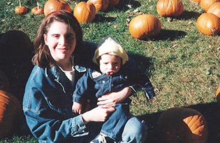
New advances in medicine and technology are helping to diminish the appearance of certain types of birthmarks in children and adults. As for my son, he is now a healthy and happy 12-year-old. His hemangioma has faded considerably to the point that it blends in with his natural skin tone, and it has regressed on its own over time.
Jennifer Lacey specializes in covering family health and lifestyle issues. She blogs at amodestmommasmusingsforlittlereaders.blogspot.com.
Read more
View all articles in the 2014 Baby & Toddler Guide